Osteoarthritis of the Elbow Patient Guide

Home
» Elbow
» Osteoarthritis of the Elbow Patient Guide
Share this page
Printer

Introduction

Physiotherapy in Barrie for Elbow Issues

Welcome to COMPHYSIO’s guide to osteoarthritis of the elbow.

The elbow joint is injured less often than many other joints in the body. The most common injuries of the elbow joint are fractures and dislocations.  Fortunately most elbow injuries tend to heal fairly well at the time of injury.

Sustaining an elbow injury, however, can lead to problems with the joint later in life.  This is true for any injury to a joint but the elbow is one of the joints particularly sensitive to developing problems later on.  The initial injury can change the way the joint works just enough to cause extra wear and tear to the surfaces of the joint. Over time, the joint degenerates, causing pain and difficulty with daily activities. This condition is called osteoarthritis (OA), degenerative arthritis, or posttraumatic arthritis.

This guide will help you understand:

  • how the condition develops
  • how health care professionals diagnose the condition
  • what can be done to ease the pain and regain elbow movement
  • what COMPHYSIO’s approach to rehabilitation is

Anatomy

What parts of the elbow are affected?

The elbow joint is made up of three bones: the humerus bone of the upper arm, and the ulna and radius bones of the forearm.

The ulna and the humerus meet at the elbow and form a hinge. This hinge allows the arm to straighten and bend. The large triceps muscle in the back of the arm attaches to the point on the end of the ulna (the olecranon). When the triceps muscle contracts, it straightens out the elbow. The biceps muscles in the front of the arm contract to bend the elbow.

The connection of the radius to the humerus and the ulna allows the forearm to rotate. The upper end of the radius is round. It turns against the ulna and the humerus as the forearm and hand turn from palm down (pronation) to palm up (supination).

In the elbow joint, the ends of the bones are covered with articular cartilage. Articular cartilage is a slick, smooth material. It protects the bone ends from friction when they rub together as the elbow moves. Articular cartilage is soft enough to act as a shock absorber.  At the same time it is also tough enough to last a lifetime, if it is not injured.

Related Document: COMPHYSIO's Guide to Elbow Anatomy

Causes

What causes OA?

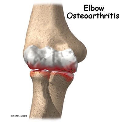
OA is caused by degeneration of the articular cartilage of a joint. Degeneration is wear that happens over time. Doctors use the term degenerative arthritis to describe the wear and tear of a joint over many years. Degenerative arthritis is another term for OA. Some doctors use the term degenerative arthrosis.  Arthrosis means that the joint is wearing out.

A bad sprain or fracture can damage the articular cartilage. The cartilage can also be bruised when too much pressure is put on the cartilage surface.  In some cases OA may be idiopathic which means there isn’t a known reason for the condition, but most of the time, elbow OA is linked with excess use of the arm or a previous injury.

At the time of injury the cartilage surface may not look any different if it has not been directly injured. The injury to the cartilage often doesn't show up until months later. In some cases, however, the damage to the cartilage with the initial injury is severe. Pieces of the cartilage can actually be ripped away from the bone and these pieces do not grow back. Usually they must be surgically removed otherwise they cause problems with the functioning of the joint. If the pieces aren't removed, they may float around in the joint, causing it to catch. These pieces are referred to as loose bodies or joint mice.  In addition to catching in the joint they can also cause a lot of pain and do more damage to the joint surfaces if left inside the joint.

Unfortunately the human body does not do a good job of repairing these holes in the cartilage surface of joints. The holes fill up with scar tissue. Scar tissue is not as slick or rubbery as the articular cartilage so the joint mechanics suffer.

An injury doesn't have to directly damage the articular cartilage to start the process of OA. Any injury to the elbow joint can change the way the joint works which then starts the cycle of wear and tear on the joint. For example, after an elbow fracture the bone fragments may not line up exactly.  If this is the case they then heal in a slightly different position from prior to the injury, which may only mildly modify how the joint works, but is enough to change the wear and tear on the cartilage.  

A dislocation can also cause lasting damage to the elbow joint. After the ligaments have been injured in a dislocation, the elbow joint may move differently. This change in movement alters the forces on the articular cartilage.  Similar to how a machine works, if one mechanism is out of balance, there can be more friction between the moving parts and it wears out faster.

Over many years, this imbalance in joint mechanics can damage the articular cartilage. Since articular cartilage cannot repair itself very well, the damage adds up. Finally, the joint can no longer compensate for the damage, and the elbow begins to hurt.


OA of the elbow is a similar process to that which occurs in the hip or knee, however, more often in the elbow, the articular cartilage itself isn’t damaged with an initial injury, whereas in the hip or knee it often is. In the elbow the biggest changes occurring after an injury are hypertrophic osteophyte (bone spur) formation and capsular contracture.

Capsular contracture refers to the process by which the capsule dries out and tightens up. The capsule has two layers: a fibrous covering that surrounds the joint and an inner lining. The inner layer is called the synovium. The synovial layer holds the lubricating fluid inside the joint.  If this fluid dries up, or the joint capsule contracts, the mechanics of the elbow joint change, and this in turn applies more pressure to the articular cartilage and wears it out quicker.

Symptoms

What symptoms does OA cause?

Pain and stiffness are the main symptoms of OA of any joint, including the elbow. At first, the pain comes only with activity. Most of the time the pain lessens while doing the activity, but after resting for several minutes pain and stiffness increase. As the condition worsens, you may feel pain even when resting. The pain may interfere with sleep.

You may have swelling around your elbow. Your elbow joint may fill with fluid and feel tight, especially after using it.  If the OA is to the stage where all the articular cartilage is worn off the joint surface, you may begin hearing a squeak and feel a creak in the joint when you move your elbow. This creaking sound or sensation is called crepitus.

OA eventually affects the elbow's motion. The elbow joint is one of the most sensitive joints in the body to injury. It quickly becomes stiff and loses motion. The first thing most people notice is that it becomes hard to completely straighten the arm. Later they find it hard to bend.


Loss of motion leads to weakness and decreased function. Carrying heavy objects at the side of the body with the elbow straight is especially difficult.

Diagnosis

How will my health care professional know if I have OA?

The diagnosis of OA of the elbow begins with an extensive history of the problem. Your health care professional will ask questions about your pain, including where exactly it is, which activities aggravate or ease it, and how your pain affects your daily activities.  Being that OA develops over a long time, you may even be asked questions about previous injuries you had or regular activities you participated in a long time ago.  For example, throwing athletes (such as baseball pitchers) have a higher risk of developing osteoarthritis of the elbow later in life. Men with a history of heavy use of the arm, such as manual laborers and weight lifters, are also at risk.

After taking a thorough history your health care professional will do a physical examination of the elbow and possibly other joints of your upper limb. It is important that they determine exactly where and when you feel your pain. Your health care professional will also feel for any catching as the joint moves. As mentioned above, loose fragments of cartilage and bone may cause this catching.

In order to help determine if OA is present you will probably need to get standard X-rays. X-rays are usually the best way to see what is happening with your bones if OA is suspected. X-rays can help your health care professional assess the damage and track how your joint changes over time. X-rays can also help your health care professional see how many bone spurs are present and if there are any loose bodies in the joint. X-rays also show the size of the joint space and how much articular cartilage is left.

A CT scan may also be ordered. CT scans can reveal osteophytes that don't appear on plain X-rays.  CT scans give a 3-D view, which will show the size and location of any bone spurs present. CT scans also show the surgeon how close any bone spurs are to the ulnar nerve of the elbow, which is important information if surgery is planned to remove the spurs.

Your doctor may order blood tests if there is any question about the cause of your OA. Blood tests can show certain systemic diseases, such as rheumatoid arthritis.

Treatment

What can be done to get rid of my pain?

Nonsurgical Treatment

In almost all cases, doctors try nonsurgical treatments first. Surgery is usually not considered until it has become impossible to control your symptoms.

Your doctor may recommend non-steroidal anti-inflammatory drugs (NSAIDs), such as aspirin and ibuprofen, to help control swelling and pain.

An injection of cortisone into the elbow joint can give temporary relief. Cortisone is a powerful anti-inflammatory medication. It can very effectively relieve pain and swelling. Its effects are temporary, usually lasting several weeks to months. There is a small risk of infection with any injection into the joint, and cortisone injections are no exception.


Finally, physiotherapy has a critical role in the non-surgical treatment of elbow OA.

Rehabilitation

What should I expect during rehabilitation for my elbow?

Nonsurgical Rehabilitation

The main goal of nonsurgical physiotherapy treatment at COMPHYSIO for OA in your elbow is to help you manage your pain, maintain your elbow range of motion and strength, and to teach you how to use your elbow without causing more harm.

During the first few appointments at COMPHYSIO your therapist will focus initially on relieving your pain.  Your therapist may use modalities such as heat, ice, ultrasound, or electrical current to assist with decreasing any pain or swelling you have around your elbow or anywhere along the forearm, at the shoulder, or into your hand. Your therapist may also use massage directly around your elbow or for your neck, shoulder, forearm, or wrist to improve circulation, assist with the pain, and make moving your entire upper extremity easier. To assist in getting rid of your pain, you may also need to modify or limit your activities that irritate the elbow. Your physiotherapist will advise you on how best to do this.

Resting the joint can help ease pain and inflammation especially if the joint is very irritable.  A relative rest, where certain irritating activities are limited but others are allowed, may be enough to calm the symptoms down. Your physiotherapist may also suggest that you use a special elbow splint to immobilize and protect the elbow.  Taping the elbow may also provide some relief.  Some patients with OA also find relief when they use a topical rub.  Your physiotherapist can advise you on if there would be any benefits to using braces, taping, creams or rubs in your individual case.

Next your physiotherapist will prescribe a series of stretching and strengthening exercises for your elbow. If you have lost any range of motion (ROM) in your ability to bend, straighten, or rotate your elbow your physiotherapist will measure this. They will also measure your grip strength on both sides.  ROM and grip strength measures are very useful in order to track improvements and to ensure that the elbow is not declining further in function.  Depending on the progression of OA in your elbow, simply not having a decline in ROM or strength may be considered very successful rehabilitation.  In other words, sometimes as long as your symptoms, ROM, and strength are not getting worse over time, even though they may not be getting better, rehabilitation can be deemed a success.

Several stretching exercises for your elbow will be prescribed. If you have lost some ROM these exercises will be used to assist in re-gaining this ROM. Usually full ROM will not be achieved if you have already lost a significant amount of motion, but improvements can most often be made. If you have not lost any ROM then the stretches are used to maintain your range, and also to maintain the length of the muscles that cross your elbow joint. Tight muscles can be detrimental to the joint as they add pressure to the articular cartilage inside.  If needed, your physiotherapist will mobilize your elbow joint. This hands-on technique is used to encourage the elbow to move into its normal ROM. With OA, mobilizing the joint can be particularly useful.  Sometimes gaining even just a few degrees of ROM allows you to use your elbow more easily and eases symptoms.

Next your physiotherapist will prescribe some strengthening exercises for your elbow. By maintaining the strength in the muscles surrounding your elbow, the pressure is minimized on the elbow joint. Strength in your wrist as well as in your shoulder and shoulder blade area are also important in order for the elbow to function optimally. The shoulder blade is the upper extremity’s link to the torso so strength and control of this area is particularly crucial in being able to stabilize the arm so that the elbow can work well.  Gripping exercises, although done with the hand, primarily use the muscles of the forearm, which connect to the elbow and are therefore an important part of your rehabilitation program. You will be pleased when your grip strength improves as daily activities such as twisting door handles or pulling or lifting heavier items such as shopping bags will become easier and less painful.  
The use of free weights or elastic bands may be incorporated into your exercise routine in order to build up the strength of the muscles supporting your elbow, shoulder, and wrist.  As your elbow becomes strong, eccentric exercises will be added. Eccentric exercises are ones where weight is applied as the muscles are lengthening.  An example of this is holding a weight in your hand and moving from a bent elbow position to a straight one. Eccentric exercises are crucial in preparing the muscles and tissues around the elbow joint to take the load of everyday activities, such as lifting a tea kettle up and then lowering it onto the stove. As you get even stronger, your physiotherapist will increase the speed of your strengthening exercises in order to further build up the strength and more closely simulate the every day activities your elbow will be required to perform.

Exercises to improve your proprioception will also be added.  Proprioception is the ability to know where your body is without looking at it.  Receptors in your joints and ligaments primarily control proprioception so an injury to the joint will have a significant impact on one’s proprioception.  Any period of immobility of the joint will add to this decline.  Even activities such as assisting yourself with your arms to get out of a chair, pulling a glass from a cupboard, lifting a pot, or pushing a door open requires weight to be transmitted through your elbow and for your body to be proprioceptively aware of your limb.  Participating in sporting activities with your upper extremity requires an even higher level of proprioceptive control. For this reason, proprioceptive exercises will be an important part of your rehabilitation regime at COMPHYSIO and your physiotherapist will be strict about your technique with these exercises in particular.  As mentioned above these exercises will also include ones for the shoulder and shoulder blade, which is the elbow’s link to the torso. Proprioceptive exercises might include activities such as rolling a ball on a surface with your hand, holding a weight up overhead while bending and straightening your elbow, or push-ups on an unstable surface. Advanced exercises may include activities such as throwing or catching a ball.  

In order to reduce your pain or assist with the biomechanics of your elbow joint, your physiotherapist may use some tape on your elbow. In some cases this can be quite useful to decrease your pain, which then allows you to strengthen the joint more easily.  The tape can also allow you to do more everyday activities without discomfort.  Some braces for the elbow can do the same thing. Your physiotherapist will advise you if taping or bracing may be useful in your situation.

We highly recommend maintaining the rest of your body’s fitness with regular exercise while you rehabilitate your elbow.  Maintaining general cardiovascular fitness can be done with lower extremity fitness activities such as walking or using a stationary bike or stepper machine.  Weights for your other arm or lower extremities can also be done but you may require a friend to assist you in the gym as lifting any weight, even just to prepare a machine could be detrimental to your elbow rehabilitation and should not be undertaken until your physiotherapist deems it appropriate.

Surgery

If non-surgical treatment is not successful, or if over time the elbow gets worse or sustains further injury, eventually, it may be necessary to consider some type of surgical treatment. There are several operations to treat advanced OA of the elbow. Your surgeon will consider many factors when deciding which procedure is best for you, including the severity of joint degeneration, your age, your activity level, and how you use your elbow.

Arthroscopic Debridement

If you are in an early stage of OA, your doctor may recommend arthroscopic debridement. Other names for this procedure include arthroscopic ulnohumeral arthroplasty or osteocapsular arthroplasty.

Arthroscopic procedures use an arthroscope, which is a tiny TV camera that is inserted into the joint through a very small incision. The arthroscope allows the surgeon to watch on a TV screen what he or she is doing inside the joint during the procedure.

In arthroscopic debridement, the surgeon makes other small incisions for inserting special tools to get rid of bone spurs, remove loose bodies, or to smooth the cartilage. Sometimes a capsular release is needed.  An incision is made in the tissue and the anterior (front) of the capsule is removed.

Your surgeon may also do lavage of the joint. Lavage involves rinsing the joint with a sterile saltwater solution, which helps remove tiny debris that may be irritating the joint.

The majority of patients treated with arthroscopic debridement for elbow OA have less pain and more motion after surgery. Symptoms may come back in some patients, but they are usually less severe.

Interposition Arthroplasty

Before the invention of high-quality artificial joints, surgeons used many techniques to keep the bone surfaces of arthritic joints from rubbing against each other. One of these techniques is distraction interposition arthroplasty. This procedure involves placing a piece of tendon or fascia between the bony surfaces of the elbow joint.  Fascia is a connective tissue, similar to tendon that lies in a flat sheet. It covers the muscles and acts as a divider between different compartments of the body.

As the joint heals, the tendon or fascia forms a cushion of thick, tough tissue between the bones. The tissue pads the ends of the bones and reduces pain while still allowing the elbow to move.

Interposition arthroplasty is rarely indicated but may be used in rare cases.  This procedure works fairly well in the elbow but doesn't work very well in the weight-bearing joints of the hip, knee, and ankle.

Elbow Fusion

A fusion surgery (also called arthrodesis) eliminates pain by making the bones of the joint grow together, or fuse, into one solid bone. Fusions were very common before the invention of artificial joints. Even today, joint fusions are commonly used in many different joints to get rid of the pain of arthritis.

An elbow fusion will greatly decrease the motion in your arm however, it does leave you with a strong and pain-free elbow. People who need a good range of motion in their elbow should consider another type of operation, such as an elbow joint replacement.

Elbow Joint Replacement

Elbow joint replacement or total elbow arthroplasty (TEA) is not nearly as common as hip, knee, or shoulder replacement. This is true for a couple of reasons. Firstly, OA in the elbow is not as common as osteoarthritis in weight-bearing joints.  Secondly, elbow joint replacement also has a higher complication rate than the more common replacement surgeries. Infection and slowed healing in the surgical incision are two complications of this type of procedure. Fracture, dislocation, and loosening are other problems reported with TEA.

Elbow joint replacement, however, is a good choice for patients who need improved motion rather than strength. Older patients who don't need as much strength will probably prefer the results of elbow replacement surgery, however, they must be willing to accept low levels of activity involving the elbow. Patients with advanced rheumatoid arthritis are also good candidates for TEA.

Post-surgical Rehabilitation

Immediately after surgery your elbow will be bandaged with a well-padded dressing and put into an elbow splint with a sling for support.  You may see a physiotherapist while in the hospital who starts you with some exercises, but this depends on what procedure you have had done and whether or not your surgeon recommends it.  If exercises are prescribed immediately post-surgery generally they are only simple range of motion exercises for your hand, fingers, and shoulder.  In some cases simple elbow exercises will also begin.  

Once you are discharged from the hospital, physiotherapy at COMPHYSIO can begin as soon as your surgeon recommends it.  Each surgeon will set his or her own specific restrictions based on what was done during surgery, personal experience, and whether the elbow is healing as expected.  Physiotherapy at COMPHYSIOmay be required for up to 3 months post-surgically, and in order to maximize rehabilitation you will need to continue your home program for an even longer period after that.

Even if extensive physiotherapy for your elbow is not immediately indicated, at COMPHYSIO we highly recommend maintaining the rest of your body’s fitness with regular exercise during the initial period after surgery.  Maintaining general cardiovascular fitness can be done with lower extremity fitness activities such as walking or using a stationary bike or stepper machine.  Weights or weight machines for your lower extremity and opposite arm are also acceptable to use as long as the restrictions regarding your healing elbow are strictly abided by.  Initially lifting even light weights with your surgical limb will not be allowed and will be difficult as your elbow heals so you may require a friend to assist you with your weights setup if you are keen to continue while your joint heals.  Your physiotherapist at COMPHYSIO can discuss the most appropriate way for you to maintain your fitness as well as provide a general fitness program specific to your needs while abiding by your surgeon’s restrictions.

Once you begin to use your elbow after surgery you will experience some pain in your elbow and forearm, and possibly even when you move your wrist or shoulder.  This is considered normal. This pain is from both not using the elbow regularly and the surgical process itself. Your physiotherapist at COMPHYSIO will focus initially on relieving your pain.  They may use modalities such as heat, ice, ultrasound, or electrical current to assist with decreasing any pain or swelling around the surgical incision, along the forearm, or into the shoulder or hand. They may also use massage for your neck, shoulder, elbow, forearm, or wrist to improve circulation, assist with the pain, and make moving the entire upper extremity easier.

As the elbow tolerates it, rehabilitation after surgery for OA will follow along similar lines of that listed under non-surgical rehabilitation. Exercises to improve and regain ROM, strength, endurance, and proprioception will be prescribed as part of an exercise program you will do in the clinic and also as part of your home exercise program.  These exercises will be advanced as your elbow heals in order to ensure that your elbow is improving as quickly as it can and that you can return to your work and recreation activities as soon as possible.  Doing your rehabilitation, exercises in a pool can be useful particularly after surgery as the warmth and hydrostatic properties of the water can often allow you to gain more range of motion with less discomfort.  Your therapist can advise you whether or not pool exercises may be useful in your situation.  In any case, following the guidance and advice of your therapist is crucial in avoiding unnecessary pain or strain on your healing elbow.

Your physiotherapist will provide hands-on treatment as needed after surgery in order to improve your range of motion and minimize your pain.  It should be noted that depending on which surgical procedure you have had done, your elbow range of motion may not improve, or may remain severely limited.  In these cases, maximizing this limited range of movement is still a goal, but a decrease in pain and an improvement in strength are the more important outcomes measured and used as indicators of a successful surgery and rehabilitation regime.

Most patients with elbow OA, whether treated non-surgically or surgically, do very well with the rehabilitation we provide at COMPHYSIO.  If, however, during rehabilitation your pain continues longer than it should or therapy is not progressing as your physiotherapist would expect, we will ask you to follow-up with your doctor or surgeon to confirm that the elbow is tolerating the rehabilitation well and to ensure that if you have had surgery, there are no hardware issues that may be impeding your recovery.

Portions of this document copyright MMG, LLC.

Share this page
Printer
Sample